Bright Ideas for Routine Medical Screening: Five Ways LED Illumination Makes Life Easier
Every hospital lab starts the day like a plate-spinner who’s had one too many coffees: dozens of slides twirling through microscopes while timers beep in perfect disharmony. One minute you’re hunting malaria parasites that have mastered the art of hide-and-seek, the next you’re coaxing faint antibody glows into confessing their secrets. Therefore, speed matters. Nobody wants a queue of clinicians asking “Is it ready yet?” – and clarity matters even more, because “sort of purple” is not a recognised diagnostic category.
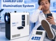
Enter LED illumination.
Unlike mercury or metal-halide lamps, which require the patience of a saint and carry a genuine risk to safety, LEDs fire up instantly, stay the same colour all day and don’t hiss ominously when you least expect it.* In short, they let you focus on the biology, not the bulb.
Below are five common screening disciplines and a look at how LEDs make each task simpler, safer and faster.
(*If your equipment does actually hiss, you’ve either got a metal-halide lamp or a very disapproving cat.)
1 — Immunology
Testing patient samples to see how the immune system is behaving – think allergy panels, auto-antibody screens or complement activity. Fluorescent markers glow when specific antibodies bind, providing a “yes/no” answer at a glance.
Why LEDs help:
-
Instant-on white light means no ten-minute warm-up while serum dries on the slide.
-
Stable colour temperature eliminates shifts that make borderline positives hard to judge.
-
Lower heat output keeps delicate fluorophores from fading mid-assay.
Metal-halide lamps, by contrast, need time to stabilise and drift in intensity over a single session exactly when you’re trying to compare subtle fluorescence from one field to the next.


2 — Bacteriology
Spotting, staining and classifying bacteria. A classic Gram stain turns Gram-positive bugs purple and Gram-negative ones pink, while fluorescent stains can highlight tuberculosis bacilli in lurid green.
LED advantages:
-
Consistent white-light illumination reveals true stain colours; no “is that purple or just warm lighting?” debates.
-
Zero mercury vapour means safer air for techs working in biosafety hoods.
-
Energy savings are substantial: LEDs sip power during the many moments when the light source is idling between samples.
Switching off a metal-halide lamp between runs isn’t an option; you’d spend all day waiting for it to re-ignite.
3 — Haematology
Counting and classifying blood cells (RBCs, WBCs, platelets) and spotting anomalies such as blast cells or malarial parasites. Bright, even illumination is key—poor light hides pale platelets and odd-shaped erythrocytes.
LED benefits:
-
Uniform field illumination prevents dark corners that could hide tiny cells.
-
Immediate full brightness lets stat samples move straight to the scope.
-
Dim-to-view control reduces eye strain during long slide reviews.
Metal-halide systems, conversely, run hot, risking slide drying artefacts and forcing techs to juggle brightness using neutral-density filters rather than a simple dial.


4 — Tissue Imaging (Histopathology & Cytology)
LExamining biopsies or cell smears for abnormal structure and staining patterns. Pathologists rely on colour consistency to judge subtle differences in H&E or immunocytochemistry slides.
Why LEDs shine:
-
Fixed, daylight-like colour temperature means slides look the same at 9 a.m. and 4 p.m.
-
Instant switching between brightfield and fluorescence channels, which is helpful when confirming a suspicious region with a quick antibody check.
-
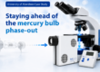
Long LED lifetimes (25 000 + hours) cut unscheduled downtime for bulb changes.
With metal-halide lamps, every change of bulb potentially alters colour balance; the pathologist notices, and the lab spends hours recalibrating cameras.
5 — Parasitology
Detecting parasites such as Plasmodium (malaria), Giardia or microfilariae in blood films and stool samples.
LED advantages:
-
Precise wavelength selection excites dyes like acridine orange without flooding the slide with extra UV.
-
Lower UV output protects techs’ eyes and reduces photobleaching, which is absolutely vital when hunting faint, moving organisms.
-
Cool running temperature keeps thick films from cracking or crystallising.
Mercury lamps produce broad UV peaks that bleach acridine orange quickly and require hefty goggles; LEDs give you the light you need, not the excess you don’t.
")
LED systems swap the lab’s traditional “brew a cup of tea while the lamp warms up” ritual for instant-on brilliance, trade wandering colour shifts for rock-steady spectra, and replace bulbs that hiss like a disgruntled kettle with quietly reliable solid-state parts.
In day-to-day screening – whether you’re chasing antibodies, bacteria, stray blood cells or an unexpectedly athletic parasite – that means quicker workflows, crisper images and fewer nervous glances at the fire extinguisher. So if your current lamp still hums, steams and occasionally performs the grand finale of Shattered Glass Concerto in D-minor, perhaps it’s time to let LEDs shoulder the load; your staff, slides and patients will thank you for the silence.
Written by Ben Furness / ben.furness@coolled.com / LinkedIn Profile