
How LED Illumination Changed Slide Scanning
Whole slide imaging has become faster, more automated and considerably more colourful. Behind all that clever scanning hardware, illumination has had a quiet little revolution of its own.
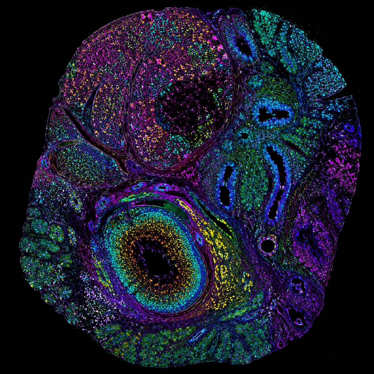
Digital pathology has come a long way from looking down a microscope, moving the slide three millimetres to the left, losing the interesting bit, then spending five minutes trying to find it again.
Whole slide imaging can now digitise complete tissue sections at high resolution, creating images that can be viewed, shared and analysed on screen. Modern systems can process large batches of slides automatically, while fluorescence scanners can capture multiple channels across enormous numbers of individual image tiles.
And somewhere behind the cameras, optics, stages and extremely clever software sits the thing making all those images visible in the first place.
From bulbs to LEDs
Traditional fluorescence microscopy relied heavily on mercury and metal halide lamps. They worked, but they came with certain personality traits.
They needed warming up. Their output changed as bulbs aged. They generated plenty of heat. Eventually the bulb needed replacing and aligning, usually at precisely the moment nobody wanted to deal with it.
LEDs behave rather differently. They provide stable output without lengthy warm-up periods, can switch electronically in microseconds and maintain much more consistent illumination throughout their operating life.
For an automated slide scanner expected to keep working through large batches of samples, that matters rather a lot.
Faster scanning, fewer wasted photons
A fluorescence slide scanner may need to capture thousands of individual images across several channels before stitching everything together into one whole slide image.
When illumination can switch rapidly between wavelengths and synchronise directly with the camera, there’s less waiting around between acquisitions. The sample also only needs illuminating while an image is actually being captured, helping reduce unnecessary exposure and photobleaching.
Rapid electronic switching reduces delays between fluorescence channels, which becomes increasingly useful when those tiny delays are repeated thousands of times.
Precise triggering means illumination can be delivered when the camera needs it, rather than bathing the sample in light while everybody waits.
Consistency matters too
Speed isn’t much use if half the slide needs scanning again.
Whole slide imaging depends on consistent image quality across a large area. Stable LED output and good illumination uniformity help maintain comparable fluorescence intensity from one field to the next, which is particularly useful when all those fields eventually need stitching together.
Consistent illumination also matters when systems are expected to run repeated scans over long periods. If the light source behaves predictably from slide one to slide one hundred, there’s a better chance the resulting images will too.
Which is generally preferable to discovering tile 2,847 looks odd.
Then multiplexing arrived
Modern digital pathology is asking scanners to capture more information from the same tissue. Rather than imaging one or two fluorescent markers, multiplexed workflows can examine many biological targets while preserving information about where they sit in relation to one another.
Multichannel LED systems are particularly well suited to this. Individual excitation wavelengths can be selected electronically, switched rapidly and controlled independently, giving automated imaging systems much greater flexibility as fluorescence panels become more complex.
A small part of a much bigger change
Better cameras, optics, automation, software and computing have all helped turn slide scanning into what it is today. LED illumination is only one part of that story.
But when you're trying to scan more slides, capture more fluorescence channels, reduce rescans and produce consistent images day after day, having a light source that switches instantly and behaves predictably is rather useful.
Sometimes progress really is just making one part of the microscope considerably less annoying.