Professor Gail McConnell’s recent presentation at the Nanoscale Optical Microscopy Meeting in Venice, was very well received. Read the abstract and download the the presentation slides below…
The Mesolens: a giant colour-corrected microscope objective lens for sub-cellular 3D resolution imaging within 118 cubic millimetres of tissue
G. McConnell, J. Trägårdh, L. McCann, R. Amor, E. Reid, J. Dempster and W.B. Amos
For more than a century, the design of low-magnification microscope objectives has been guided by the angular acuity of the human eye (approximately 1 second of arc). At x4 magnification, this requires a numerical aperture no greater than 0.1 or 0.2, which can be achieved cheaply and easily by simple optical designs. With the advent of confocal and multiphoton microscopy, however, it became apparent that the poor axial resolution of more than 30 microns with low magnification objectives was intolerable for these 3D methods.
To overcome this, we have developed a new and complex objective with a magnification of 4x and an NA of just less than 0.5 which we call the Mesolens. We specified this lens for mammalian embryology, and have shown that it can image every cell of a 6mm-long embryo 3mm thick with sub-cellular resolution if the tissue is cleared appropriately. A by-product of the high NA is that the optical throughput is approximately 20x greater than a conventional 4x objective. The pupil size of the lens is so great that it cannot be used with a conventional microscope frame, so we have built the imaging system around the lens, and use either wide-field camera or point-scanning fluorescence detection to create images. Applications in neurobiology and many other branches of biomedical science and beyond are likely.

Above: Confocal laser scanning Mesolens dataset showing in a single image almost half of a mouse brain section, stained to show as bright dots aggregates of SOD1, a protein which shows a prion-like misfolding which spreads through the brain and causes amylotrophic lateral sclerosis (Huntington’s Disease). Scale bar = 1 mm. Right: An expanded region of image on the left reveals hundreds of aggregates of SOD1. Scale bar = 1 mm. (Unpublished work, specimen courtesy of Dr Anne Bertolotti, MRC Laboratory of Molecular Biology, Cambridge, UK).
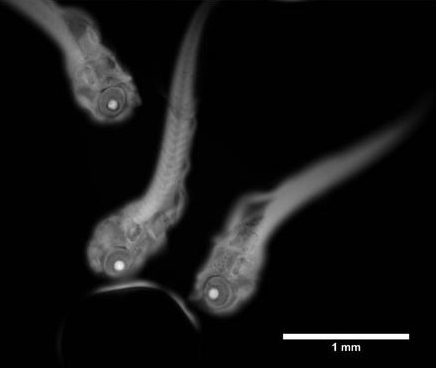
Zebrafish larvae, one day after hatching. Specimens were fixed and stained with acridine orange, then imaged with 20 ms camera exposure in widefield epi-fluorescence mode using a CoolLED pE-4000 illuminator
DOWNLOAD PROFESSOR GAIL McCONNELL’s PRESENTATION SLIDES