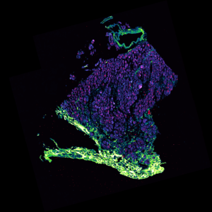
Fish Head Image in an Image by Dr Sam Duwe
We had some stunning entries for this year’s Image in an Image competition, where we look for what’s in an image, and not just the quality of the acquisition itself. The prize is one of our most popular Illumination System – the highly controllable pE-300ultra, which offers intense, broad-spectrum LED illumination for imaging most common fluorophores.
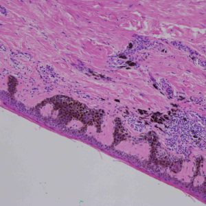
The competition was a close call, with ‘Fish Head’ by Dr Sam Duwe (Advanced Optical Microscopy Centre, Hasselt University, Belgium) coming first place in the voting. This was followed by ‘Animals in the Skin’ by Dr Charlie Tilley (NHS, and submitted by the Biomedical Imaging Unit, University of Southampton and University Hospital Southampton NHS Foundation Trust):

Animals in the Skin by Dr Charlie Tilley
About the winning image & winner
The ‘Fish Head’ image was acquired using a combination of two-photon excitation fluorescence microscopy and SHG microscopy on an unstained tissue sample. The total image size is 1.85×1.85 mm, acquired as a tilescan image with a 20x/0.8NA objective on a confocal microscope. Dr Duwe comments:
“The moment I acquired the image I immediately saw a fish head instead of a tissue sample. As the core facility manager and imaging specialist I am always looking for ways to improve my user’s imaging experience. I’m a big fan of LED Illumination Systems instead of the typical mercury/metal halide lamps even if it’s just to save on warm-up and cool-down waiting times. So this CoolLED pE-300ultra will definitely be used to replace one of those.”

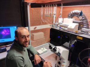
Dr Sam Duwe, winner of CoolLED’s Image in an Image Competition 2021
We will be running the competition again next year. If you want to be in with the chance of winning a 3-channel pE-300ultra LED Microscope Illumination System, make sure you sign up to our newsletter or follow us on LinkedIn or Twitter so you hear when to submit your entries for the Image in an Image Competition 2022!


