
More than a gut feeling: Revealing how microbes respond to stress
The human gut is home to trillions of microbes, which, to put that into perspective, amounts to a biomass roughly comparable to a small adult hamster. These bacteria are not just along for the ride, but play key roles in digestion, immunity and overall health. However, what happens when conditions change and nutrients run low? Understanding what happens to the molecular machinery at the single-cell level is a step towards explaining a range of gut-related issues such as inflammatory disorders. In a fascinating study published in Communications Biology, Liu et al. used a combined imaging and sequencing approach to show just how differently two gut bacteria respond when conditions start to shift.
As you would expect, life in the gut is anything but steady and bacteria must adjust quickly to survive. But because many gut microbes are anaerobic and do not behave well outside their native environment, they can be tricky to study in culture. This is why many studies utilise sequencing data, but as we know this provides limited insights into the true dynamic nature of an individual living cell. Here, the authors combined fluorescence microscopy with a variety of labelled molecules and RNA-seq to track how Bacteroides thetaiotaomicron and Roseburia intestinalis changed across growth stages and in co-culture.
Extra insights from microscopy
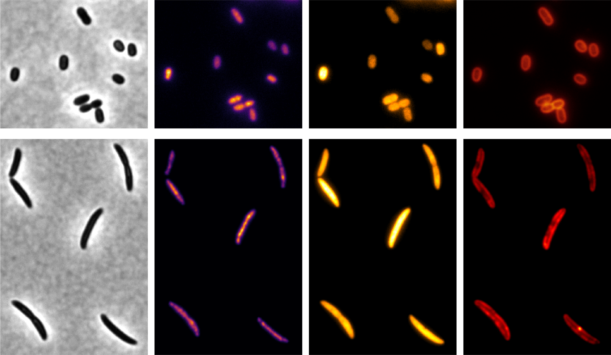
The authors used fluorescence imaging to simultaneously visualise DNA, RNA, membranes and new peptidoglycan synthesis of the cell wall, which gave them a much richer picture of cell organisation than sequencing alone could provide
| Molecule | Label | pE-800 LED (nm) |
|---|---|---|
| DNA | Hoechst 33343 | 400 |
| Bacterial peptidoglycan synthesis (the cell wall) |
HADA | 400 |
| RNA | RNASelect | 470 |
| Bacterial cell membrane | FM4-64 | 580 |
A flexible multiwavelength Illumination System such as the CoolLED pE-800 is ideal for this kind of work because it supports fast switching between individual excitation channels, stable illumination and clean separation of fluorophores, all useful when you want to follow several labelled structures in the same anaerobic sample.
The setup made use of the ability to fit single-band filters inside the light source itself, to create a Pinkel-style configuration, combining single-band excitation with multi-band dichroic and emission filters. In this case, an additional single-band filter wheel added extra specificity. Together, this equipped the researchers with the speed needed for dynamic live cell imaging, and the specificity to accurately separate and therefore quantify four molecular markers.
The light source helped move the study from mapping gene expression patterns to understanding what the cells are really doing in terms of visible changes in morphology, division and intracellular organisation.
So what did they find out?
Cell behaviours & interactions
The two species behaved very differently. B. thetaiotaomicron followed a more conventional growth pattern compared to R. intestinalis, with cell constriction and division tightly coordinated. When nutrients ran out, B. thetaiotaomicron cells became wider, whereas R. intestinalis cells became shorter and showed stronger signs of restructuring as a result of stress.
That difference matters because it suggests gut bacteria are not just passively surviving starvation, but actively remodelling their cell biology in different ways. The co-culture experiments were especially interesting: growing the two bacteria together reduced some of the nutrient stress-associated shape and transcription changes.
Gut bacteria do not all cope with stress in the same way, and those differences depend on both growth stage and who else is in the neighbourhood.
Beyond this scientific discovery, this research is exciting in that it demonstrates an approach for using fluorescence microscopy and sequencing as complementary techniques to uncover microbial behaviour at the single-cell level.