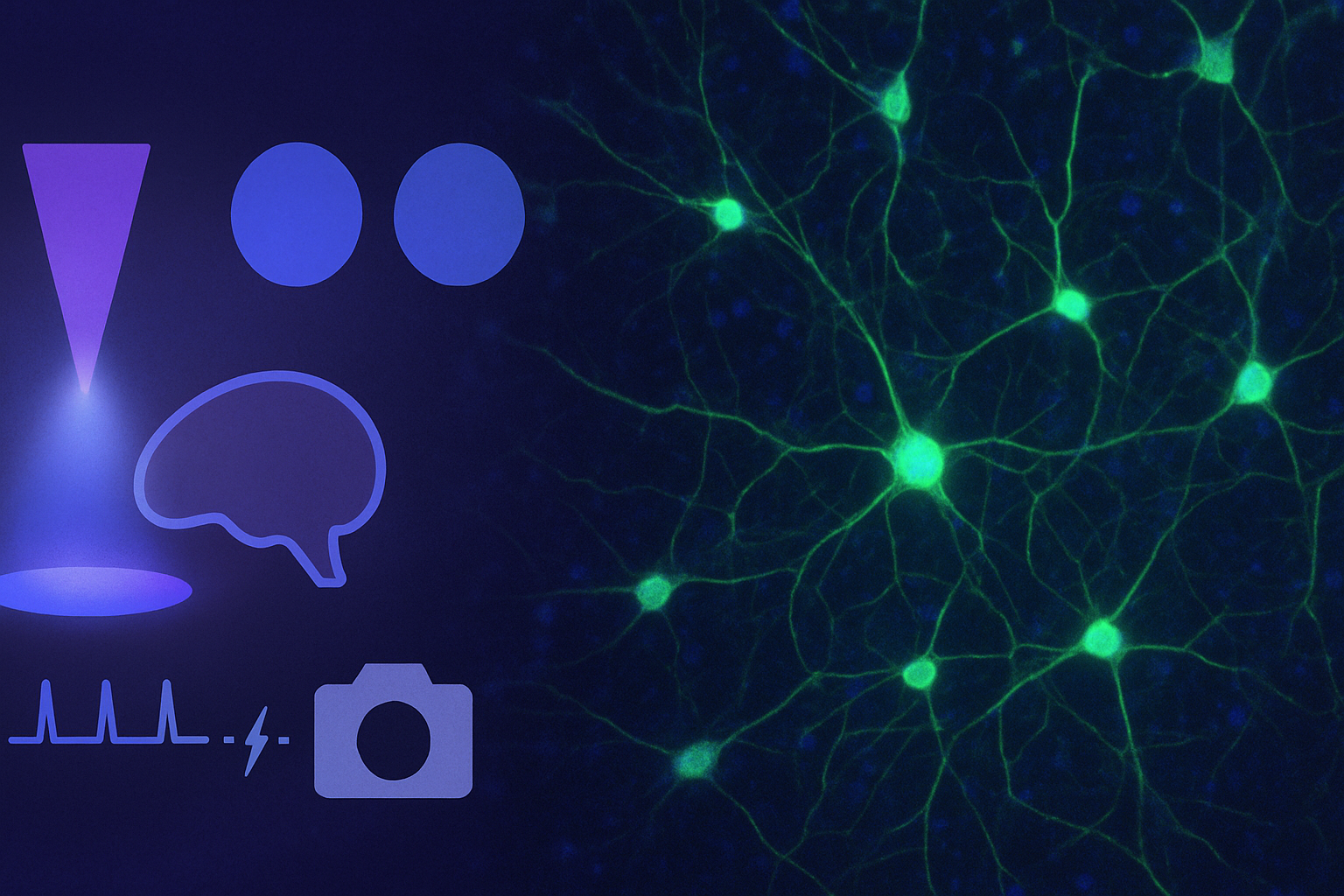
LED Illumination for Neuroscience
Neuroscience pushes light to do tricky things: excite fluorophores without cooking brain slices, toggle opsins without jitter, and keep signals flat for long recordings.
That’s why LED illumination is a natural fit – instant on, low heat at the sample plane, and rock-steady output make it easier to get clean data on the first try.
Below is a practical tour of common neuroscience workflows – calcium imaging, optogenetics, brain slices, in vivo widefield and patch-clamp-friendly setups
Why LEDs make sense in neuroscience labs
-
Instant readiness: No lamp warm-up or re-strike delays; perfect for brief trials and unplanned repeats.
-
Lower heat, less drift: Cooler optics and stable spectra help long recordings and delicate tissue.
-
Clean timing: Microsecond-scale electronic switching tracks camera exposure; less “light when you don’t need it.”
-
Mercury-free, low maintenance: Fewer interruptions, fewer consumables, fewer forms.
- Lightning fast TTL switching to help avoid photobleaching or phototoxicity
Calcium imaging (Fura-2, GCaMP & friends)
Fura-2 needs precise 340/380 nm alternation and good timing. A dual-wavelength LED makes this straightforward: set two bands, sync to the camera, and let the software compute ratios.
(Purpose-built: pE-340fura; multi-channel with 340/380 configs: pE-800fura.)
For GCaMP/red sensors, the priorities are uniform fields, stable intensity and minimal dose for long movies. Multi-band systems help when you add structural markers or a second indicator.
(Flexible all-rounder: pE-4000.)
Tip: Gate the LED from the camera’s TTL so light only appears during exposure—lower bleaching, cleaner ΔF/F.

Optogenetics
Driving ChR2, eNpHR or red-shifted opsins is about matching wavelength and controlling pulse timing. LEDs deliver repeatable flashes without shutters or filter wheels, and you can interleave stimuli with imaging frames.
If you juggle several opsins, a wide selectable spectrum keeps options open; for simpler widefield stimulation alongside imaging, bright, compact white systems are handy.
(Broad palette: pE-4000; neat white sources: pE-400max, pE-300ultra.)
Tip: Plan stimulation and imaging spectra together so opsin activation doesn’t bleed into your indicator channel.

Acute brain slices & in vivo widefield
Slices demand stable intensity and low heat; LEDs help both, which is kind to patch recordings and long protocols. In in vivo widefield, uniform illumination and instant on/off reduce fiddling between trials.
If you swap frequently between vessel labels, GCaMP and behaviour-linked recordings, quick access to multiple bands saves time. (Multi-band for variety; bright white for reflected-light context.)
Tip: Keep trigger cables short and tidy to minimise electrical noise around sensitive amplifiers.

Synchronisation: small tweak, big payoff
Treat the LED as a silent shutter. With TTL/trigger control, the light is on only when the sensor is integrating. Benefits: lower dose, steadier baselines, easier closed-loop timing, and tidy multi-colour sequences (blue → green → red) without touching a filter wheel.
Final thought
Neuroscience mixes fragile tissue, fast events and long timelines. LED illumination removes a lot of friction. No warm-ups, no drifting spectra, no mechanical shutters, and adds what you need: clean timing, stable output and sensible wavelength control.
Whether you’re quantifying Ca²⁺ with Fura-2, mapping cortical dynamics with GCaMP, or pairing stimulation and imaging in an optogenetics paradigm, the right LED setup shortens the path from preparation to publishable data and keeps the experiment (and the experimenter) that little bit calmer.
Written by Ben Furness / [email protected] / LinkedIn Profile


