Hooked on LED Microscopy Illumination After Winning a pE-300ultra
Winner of Image in an Image competition 2021 shares ‘Fish Head’ discovery and benefits of new LED Illuminator(s)
We recently spoke with last year’s Image in an Image Competition winner – Sam Duwé, Facility Manager at the Advanced Optical Microscopy Centre, Hasselt University in Belgium.
Tell me about your role:
I run a core facility here at the Biomedical Research Institute of the Hasselt University in Belgium. While we are located at the Biomedical Research Institute, we also cover researchers from areas like materials sciences. The facility has a range of techniques available, including single molecule imaging, spectral detection, correlation spectroscopy and regular widefield microscopy. I started working here at the end of 2019, which was also the time that the facility was officially founded. The equipment has always been here, but at this point we started to follow a core facility model and I’m here to help the researchers with imaging and getting the best out of their data. During my PhD and postdoc, people would always come by and ask for advice and it was interesting to look into what they were doing and how I could help. So I enjoy the variety of methods and samples and helping scientists improve their skills – that combination works perfectly for me in the core facility role.

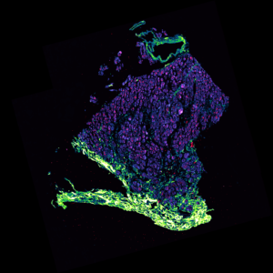
Tell us about the ‘Fish Head’ competition entry
I think I acquired the image one or two days before I saw the announcement for the competition, so it was really fresh in my head. I was lucky because the fish head appeared on screen exactly as you see it – if it had been upside down, I might have missed it. And then I figured if I’m participating in the competition I’ll make the colours stand out. I thought it looked like a real sea monster!
How has the pE-300ultra benefited your facility?
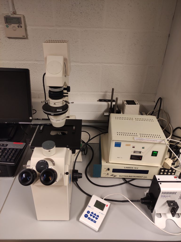
The pE-300ultra has already been used on multiple setups. The first setup I selected was an older inverted Zeiss microscope which was heavily underused because it no longer had a fluorescence light source. By coupling the pE-300ultra, we basically had a new system that even enabled us to perform live cell imaging. Then some researchers came to me with a dye that excited really well under low wavelengths. Since this was perfect for the 365 nm LED in the pE-300ultra, I moved it onto another microscope which had previously been fitted with an HBO lamp.
I’m now advising everyone to get one of these pE-300 Series Illumination Systems – it’s easy to move around, you can plug it into different systems, and there is basically no maintenance on it whatsoever. It’s also very simple to use. You just switch it on and use the control pod to control the irradiance and you’re good to go! No worrying about warming up and cooling down times, it’s just plug and play. That’s the wonderful thing about it.


You also recently purchased a pE-800 for your facility – what drew you to this product?
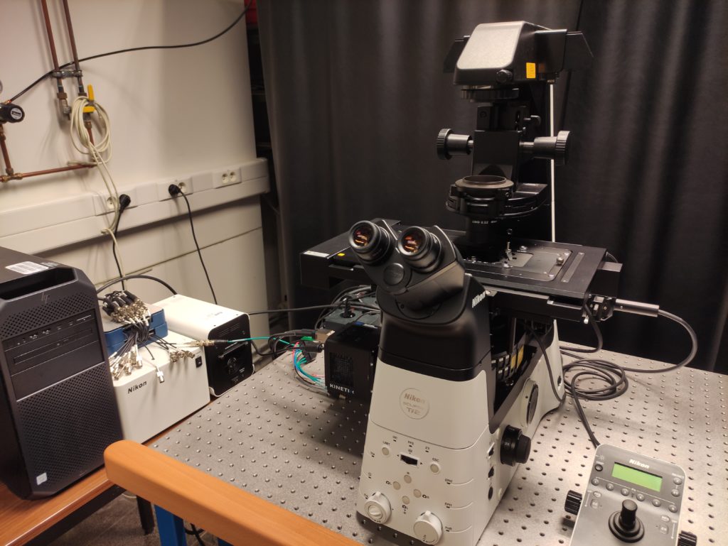
We recently purchased a Nikon widefield microscope with a large field of view and this Illumination System was a good fit. We checked the specifications of a few LED illuminators from different manufacturers and the pE-800 was exactly what we needed. I wanted a full spectrum of excitation wavelengths from the UV to the near infrared, with a good range of wavelengths in the green.
I get a lot of visitors from different labs and they all have their own preferred fluorophores. If you only have one excitation band around that area, they can’t achieve optimal excitation. Now, with this set up, they can. We also wanted our set-up to be equipped with a full high-speed TTL triggering interface for live cell imaging which we plan to start using soon.
Did anything surprise you about LEDs?
I’m always surprised at the brightness of LED light sources these days. Most people think that the brighter the excitation light is, the brighter the signal will be, and they are used to setting the irradiance to 25, 50 or even 100%. But for both the pE-300ultra and the pE-800, I constantly need to tell them to start low at 1 or 2%, which will produce plenty of signal, and you can always increase this.
But if you start at a very high irradiance, the sample will be saturated anyway and you have no idea how fast the fluorescence could bleach – you can have the best fluorophores available and they’ll still bleach. So that’s one of the things that surprised me – the huge available power.


Do you have any advice for entrants to this year’s competition?
I’d say go for colour! Add some nice colours to the image and you’ll stand out. Not just your usual red/green/blue, but go for a wacky look up table with some interesting colours – especially if it fits the theme of your image.
For more information on the core facility, please visit the Advanced Optical Microscopy Centre website.

