Automation on a budget
When setting out to capture fast cellular processes with live cell imaging, it has historically been the case that widefield microscopes were unable to achieve highspeed multi-labelled fluorescence imaging without the addition of motorised components. In these configurations, the light source has often been overlooked as an affordable means of achieving multi-wavelength imaging – but LEDs have changed the game.
In this document, we examine and compare typical configurations for automated microscope illumination systems in terms of speed, cost and signal-to-noise ratio. In particular, we explore where users can capitalise on the switching speeds of LEDs to achieve fast wavelength selection with lower cost filter sets and minimal requirement for expensive motorised components.
Find out:
• Why automated widefield microscopy no longer depends on motorised components
• Which microscope configurations prioritise speed, contrast and budget
• How to capitalise on the switching speeds of LED illumination systems
Typical configuration for achieving automated multi-wavelength imaging
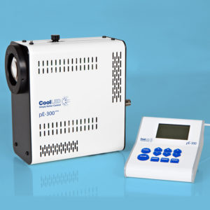
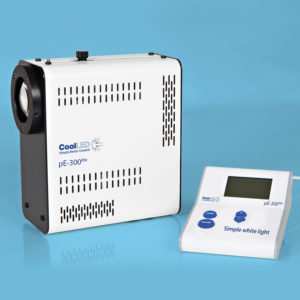
Many widefield fluorescence microscope systems include white light sources, such as mercury or metal halide lamps, and more recently “white” LED illumination systems (such as the CoolLED pE-300lite). Individual LEDs emit a discrete, relatively narrow spectrum of light, but combining LEDs of different wavelengths produces a broad spectrum white light. In such configurations, selecting a wavelength corresponding to each fluorophore requires optical filters in the excitation and emission paths. Automated wavelength selection is usually achieved with single-bandpass filter sets mounted in filter cubes within a motorised microscope filter cube turret. This approach performs well in terms of signal-to-noise and therefore image quality, since the excitation and emission is controlled with filters precisely designed for each fluorophore.
However, microscope filter turrets have relatively long switching times, limiting dynamic observations of multi-labelled samples to the order of seconds. When upgrading a manual microscope, this also relies on costly investment in multiple filters and access to a microscope which supports a motorised filter turret.
High-speed automated multiwavelength imaging with filter wheel and white light source
To overcome the speed limitations of the motorised filter turret, faster observation with a white light source can be achieved by using Pinkel or Sedat filter sets loaded into highspeed external filter wheels.
• Pinkel filter set: single-band excitation filter; multi-band emission filter and dichroic mirror
• Sedat filter set: single-band excitation and emission filters; multi-band dichroic mirror
Whilst a Sedat filter set provides the highest signal-to-noise ratio, it does require extra filters and an extra filter wheel.1 Filter wheels available at lower price points can take up to 100 ms to rotate to a new filter position, however more expensive filter wheels can offer switching speeds in the region of 55 ms with synchronisation. While this may seem to be a fast solution at first glance, filter wheels do create mechanical vibrations which may last for hundreds of milliseconds. These pass through the microscope frame resulting in image degradation, and to eliminate vibration artefacts the observation rates are often reduced to allow for vibrations to settle before image capture.
Controllable LED illumination systems for high-speed multi-wavelength imaging
Being solid-state, LEDs can be switched on and off electronically at exceptionally high speeds. More advanced LED illumination systems take advantage of this by including the ability to individually control each LED. As we will explain,this technology has the potential to provide the best of all three core aspects for multi-wavelength imaging, achieving:
• High-speed
• Low system costs
• High signal-to-noise ratio
When using an illumination system with individual channel selection such as the pE-300white with full multi-band filter sets, any latency associated with motorisation is removed. This is a low cost approach which allows multi-labelled samples to be observed with high switching speeds and without the requirement for motorised components.
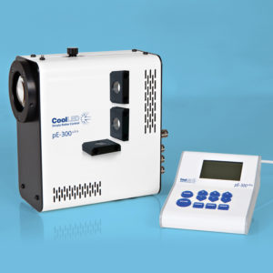
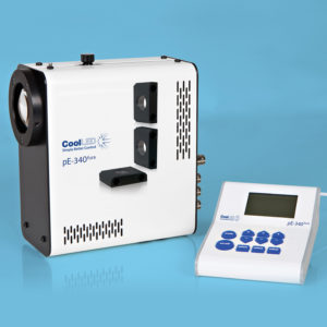
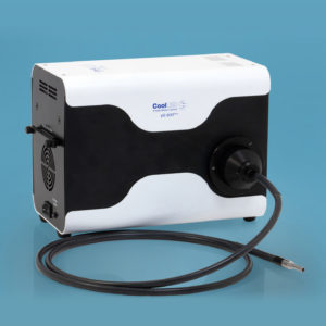
The trade-off with multi-band filters can sometimes be bleedthrough and a reduction in the signal-to-noise ratio if optical filters for multi-labelled images are not chosen carefully. To overcome this and still maintain speed and cost benefits, some LED illumination systems (such as the pE-300ultra, pE-340fura and pE-800) include inline filter holders. These allow users to incorporate individual excitation filters (from a Pinkel filter set) to further improve the signal-to-noise (Figure 1), without the cost or latency of filter wheels. Although the increased signalto-noise ratio of a Sedat filter set is not possible here, the slight compromise in image quality comes at the gain of significant speeds at a much lower price point.

Taking speed one step further
Whilst LEDs have the potential to offer high switching speeds, as with any peripheral device, latencies can be introduced by USB serial communications and computer operating system overheads. This can reduce switching speeds by as much as 100 ms and may also impact system synchronisation.
For users wishing to capitalise on the maximum speed of LEDs, we recommend electronic control via TTL which offers switching speeds of around 10 µs, and even down to 7 μs with the pE-800. The trade-off here is cost, and TTL control often requires specialist computer cards and components which can be expensive.
To overcome this and make TTL a cost-effective means of achieving high-speed imaging, we developed the Sequence Runner mode in the pE-300ultra, pE-340fura and pE-800 LED Illumination Systems. Sequence Runner uses the TTL output signal available on most scientific cameras and cycles though LEDs in a user-selected sequence for each TTL signal. This is a simple approach and provides high-speed microsecond switching speeds that are precisely synchronised to the camera exposure.
Limiting unnecessary sample irradiance taking place outside of the camera exposure time (which is also known as “illumination overhead”) minimises phototoxicity and photobleaching. With the resulting improvement in cell viability, this achieves not only insightful but accurate data.2
Quick comparison
The table below summarises the benefits, drawbacks and component requirements of the four configurations discussed, using an example of an automated three-wavelength system (e.g. DAPI, FITC, TRITC).

Conclusion
Many configurations are possible when seeking the optimal balance between speed, cost and contrast. However, reaching the highest temporal resolution requires a controllable LED Illumination System which can achieve switching speeds of 10 μs. These speeds may be achieved with a full multi-band filter set which also saves costs over traditional motorised filter wheels and turrets. Alternatively, overcoming the drawbacks of bleed-through and achieving a high signal-to-noise ratio, we recommend individually switchable LED illumination systems which also feature inline single band filters.
Sequence Runner further simplifies individual channel control and reduces cost, with the option to set up multi-colour highspeed imaging with high signal-to-noise via a single TTL. Not only does this present a new approach for increasing temporal resolution, but the tight synchronisation has the added benefit of enhancing data accuracy through minimised phototoxicity
and photobleaching. The light source might seem like an unlikely enabler for highspeed imaging, but the level of control that comes with solidstate LEDs is a world away from traditional mercury and metal halide lamps.
If you have any questions or would like to know more about the configurations explored in this article, please contact your local CoolLED reseller or Field Sales Manager, or simply contact us at info@coolled.com.
References
1. Erdogan, T. (2006). New optical filters improve high-speed multicolor fluorescence imaging. Biophotonics International. 13. 34-39.
2. Kiepas, A., et al. (2020). Optimizing live-cell fluorescence imaging conditions to minimize phototoxicity. Journal of cell science, 133(4), jcs242834. https://doi.org/10.1242/jcs.242834