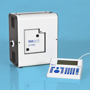
Our group is currently attempting to isolate and stain circulating tumour cells. In order to identify these cells we need a minimum of four colour fluorescence imaging, requiring the slide to be scanned using four different LEDs. To stitch our images together after scanning we require a transmission light microscope image. The image co-ordinates of the light microscope image are used to stitch the fluorescence images together. The previous light source (pE-2) allowed only three LEDs to be used together for our purposes. This meant that when we have multiple slides there are two options to get the full four fluorescence scan:1) Change the LED after scanning in Brightfield and three fluorescence channels – this risks damage to the LED itself and is time consuming. It involves un-screwing a panel each time and screws can be damaged or lost relatively easily. It places demand on the ribbon that is connected to the LED and, over time, may damage the ribbon also. We usually scan multiple slides and prefer to use option two, to reduce wear and tear on the light source.2) This option is to scan all slides in Brightfield and three colour fluorescence and then go for a second batch of images in both Brightfield and the final fluorescence channel. This is also time consuming, as setting up each scan in focus takes time. It also requires a lot more downstream processing to line the images up with one another, as the scans are often of two different sizes. With the new pE-4000 light source I was able to scan four slides in Brightfield and four fluorescence channels in three hours. Four slides took six hours to scan on the previous light source, due to double the number of scans and changing the LED. It has also reduced the downstream processing of the images, as they are all taken using the same co-ordinates, in one batch. Our project aims to expand from four colour fluorescence to six or seven channels over the coming years. Using the old light sources this would require a minimum of three but possibly four scans taken from different positions per slide and would take hours per slide. The current light source meets the needs of researches using modern immunofluorescence techniques as most projects have moved from single to a multi-marker approach, in recent years.
Anthony Cooney, PhD Student, Department of Histopathology, Trinity College Dublin (pE-4000)
Need something a little different?
For manufacturers who wish to gain a competitive advantage through bright, stable and controllable illumination, CoolLED’s world-renowned LED technology is available in customised configurations.
- Streamlined development using proven off-the-shelf technology
- Professional support
- Market-leading warranty
- ‘Try before you buy’ option
- Trusted experts in LED illumination technology since 2006

Sustainability
Did you know that LED illumination systems help laboratories reduce their carbon footprint, protecting laboratory staff along the way?
Toxic mercury-based microscope illuminators are bad news for the environment, draw a lot of power and have a short lifetime.
LED illumination systems are cleaner, have a long lifetime and use less power. Our pE-300 Series is also ACT label certified and is a natural choice for labs who want to play their part in helping the environment.
Help & Advice
At CoolLED we are committed to giving our customers outstanding service right from the start and throughout the lifetime of the product. Please get in touch if you have any questions or would like a quote. You can also browse our site for technical resources and general support.
