Vasopressin and oxytocin excite BNST neurons via oxytocin receptors, which reduce anxious arousal
Authors
Walter Francesconi, Valentina Olivera-Pasilio, Fulvia Berton, Susan L Olson, Rachel Chudoba, Lorena M Monroy, Quirin Krabichler, Valery Grinevich, Joanna Dabrowska
Topic
Neuroscience
Extract
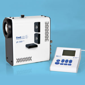
"To visualize OTR- and CRF-mCherry fluorescent neurons in brain slices from OTR-Cre and CRF-Cre rats or AVP-eYFP neurons in the hypothalamus (SON, SCN, and the PVN), we used an upright microscope (Scientifica Slice Scope Pro 1000 fitted with fluorescent filters (49008_Olympus BX2_Mounted, ET mCherry, Texas Red, ET560/40x ET630/75m T585lpxr for the visualization of mCherry and 49002 ET-EFP (FITC/Cy2) ET470/40x ET525/50m T495LPXR for the visualization of eYFP) and infrared differential interference contrast [IR-DIC] optics), with a CoolLED pE-300ultra, broad-spectrum LED illumination system as the light source.
This TLS protocol was used in BNST slices from AVP-Cre rats injected with Cre-dependent AAV-ChR2 in the hypothalamus and was delivered at the recording site using whole-field illumination through a 40x water-immersion objective (Olympus, Tokyo, Japan) with a pE-300ultra CoolLED illumination system (CoolLED Ltd., Andover, UK). "
Product Type
Journal
Cell Reports
Year of Publication
2025