white
Authors
Koha et al.
Topic
Cell Biology
Extract
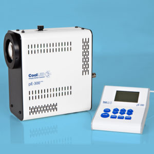
"With the Nikon microscope, red and green fluorescence were captured using a pE-300white CoolLED filtered by Nikon FITC B-2E/C and TRITC G-2E/C filter cubes, respectively. With the Zeiss microscope, they were captured using a pE-4000 CoolLED filtered by Zeiss 38HE and 43HE filter cubes, respectively. To achieve optimal image resolution without excessive illumination, a binning factor of 2 by 2 for the sCMOS camera and 5 by 5 for the CCD camera was applied prior to imaging. Throughout each experiment, the ambient conditions of the imaging platform (e.g. external lighting) were maintained to minimise variations in optical resolution and illumination"
Product Type
Journal
Journal of Cell Science
Year of Publication
2016