Authors
Larocque et al.
Topic
Medical Research, Molecular Biology
Extract
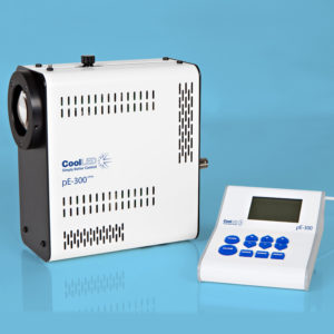
"Cells in CDM were imaged for 16 h using an Eclipse Ti inverted microscope (Nikon) with a 20×/0.45 SPlan Fluar objective and the Nikon filter sets for bright field and a pE-300 LED (CoolLED) fluorescent light source with imaging software NIS Elements AR.5.20.02.
"
Product Type
Journal
JCB
Year of Publication
2021

