Authors
Whitney J Walker, Kirsten L Underwood, Patrick I Garrett, Kathryn B Lorbacher, Shannon M Linch, Thomas P Rynes, Chloe Sloop, Karen Mruk
Topic
Cell Biology, Neuroscience, Time-Lapse Microscopy
Extract
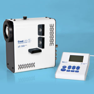
"Larvae were then washed 3 times for 5 minutes each in fresh E2 buffer, mounted on glass slides in 1% LMP agarose, and imaged on a Leica DM6D epifluorescence microscope with a Leica DFC900GT camera and CoolLED pE-300 Ultra light source or on a Zeiss LSM 700 confocal microscope equipped with a MA-PMT with 20x/NA 0.5 water immersion objective.
Following behavior experiments, larvae were imaged and scored for a cellular bridge by an independent experimenter using Leica DM6D epifluorescence microscope with a Leica K8 camera and CoolLED pE-300 Ultra light source."
Product Type
Journal
Developmental Biology
Year of Publication
2025