Dr Debbie Wilkinson – Senior Microscopy Application Specialist at The Microscopy and Histology Core Facility, University of Aberdeen (Scotland).
The Microscopy and Histology Core Facility at the University of Aberdeen supports a variety of research groups, as well as external customers, which means we have a wide range of equipment to work with diverse sample types. The Marine Laboratory brings us whale blubber and seawater, and other samples include nanoparticles, yeast, bees and mites, as well as standard mammalian cells. We also work on developing techniques, such as correlative microscopy using our fluorescence microscopes.
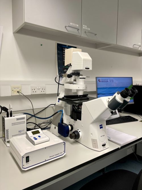
Like many imaging facilities, we have relied on traditional lamps for fluorescence microscopy illumination. However, when the bulb of the spinning disk confocal system failed, Alan Tilley (Atlantic Imaging) supplied a CoolLED pE-300lite Illumination System as a temporary measure. The Illumination System became popular as users appreciated its ease of use, with simple on/off switching and 0-100% irradiance control. Not needing to change the bulb is also convenient for those of us working at the facility. We therefore decided to replace the lamp on our widefield fluorescence inverted microscope with the pE-300lite (Figure 1).

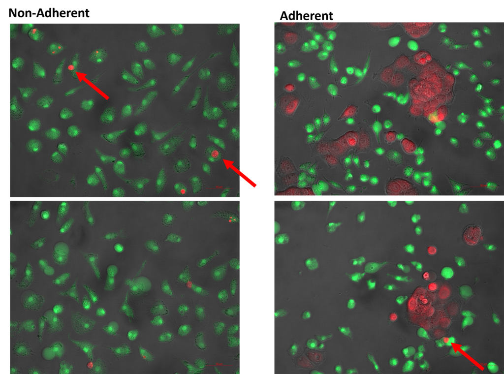
Tatties is a very heavily used workhorse system for everyday applications, for example checking sample staining before applying other techniques such as confocal laser scanning microscopy. One user is PhD student Ellen Main, who is studying how to re-programme the innate immune system for treating malignancy – specifically targeting ovarian cancer. The main aim of her project is to enhance macrophage phagocytosis of ovarian cancer cells using an antibody to cancer-associated antigens. As part of this, she has been developing methods to determine the optimum conditions for antibody binding, and assessing phagocytosis across different conditions as shown in Figure 2. CD14+ monocytes are first isolated from peripheral blood and cultured for five to seven days before the macrophages are then used for phagocytosis assays.

The facility has several fluorescence microscopes, and we may end up replacing the other bulbs in the future with LED microscope lighting. Based on our recommendation, at least two other researchers in the University have since switched to CoolLED Illumination Systems on their own microscopes.
Find out more
*The microscope is named “Tatties”, which is a Scottish term for potatoes!
