![]() Dr Darren Thomson (Experimental Officer at the Manchester Fungal Infection Group, UoM)
Dr Darren Thomson (Experimental Officer at the Manchester Fungal Infection Group, UoM)
The Manchester Fungal Infection Group (MFIG) was created in 2013 within the University of Manchester to understand and reduce the increasing burden of human fungal disease in the developed and developing world. A key technology employed by MFIG in understanding disease is our live-cell imaging suite. Our Director Prof. Nick Read was recruited from Edinburgh to Manchester along with his imaging equipment. This included Nikon wide-field acquisition systems with associated CoolLED pE-100, pE-1 and pE-2 units. These units have been reliably used by Prof. Read’s group and MFIG for >11 years now to publish live-cell pathogen and host cell biology in high-impact journals. When I arrived to MFIG and heard how long these LEDs have been used, I was a little concerned that they might be on their last legs. However, they have shown no signs of power loss, are hassle-free and will remain an integral part of our imaging suite.
We regularly perform assays which look at both the host and pathogen, where keeping both alive and happy is no mean feat. Luckily, we can employ low-power, fast-trigger excitation LEDs with our pE-Series Illumination Systems to perform live cell imaging, involving calcium signalling, fluorescent anti-microbial peptide dynamics and the fungal/host cell biology therein at high temporal frame rates.
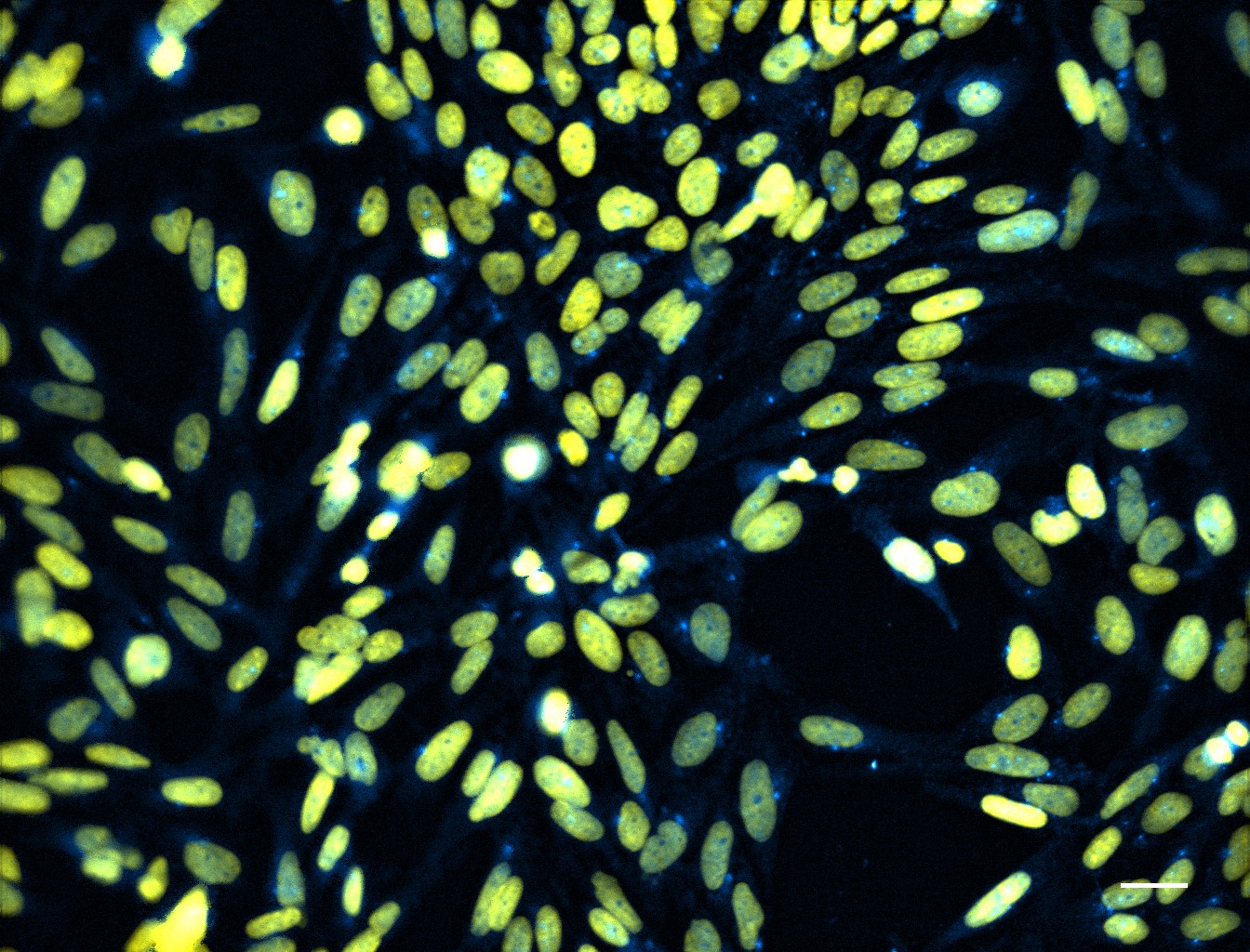 Huwe1 protein localisation, via FITC antibody labelling (blue), with respect to the DAPI stained nucleus (yellow), in human 16HBE bronchial epithelial cells. Scale bar = 10 µm. These experiments focussed on understanding host genetic factors which are important for the development of fungal disease. (Image courtesy of Dr Sara Gago).
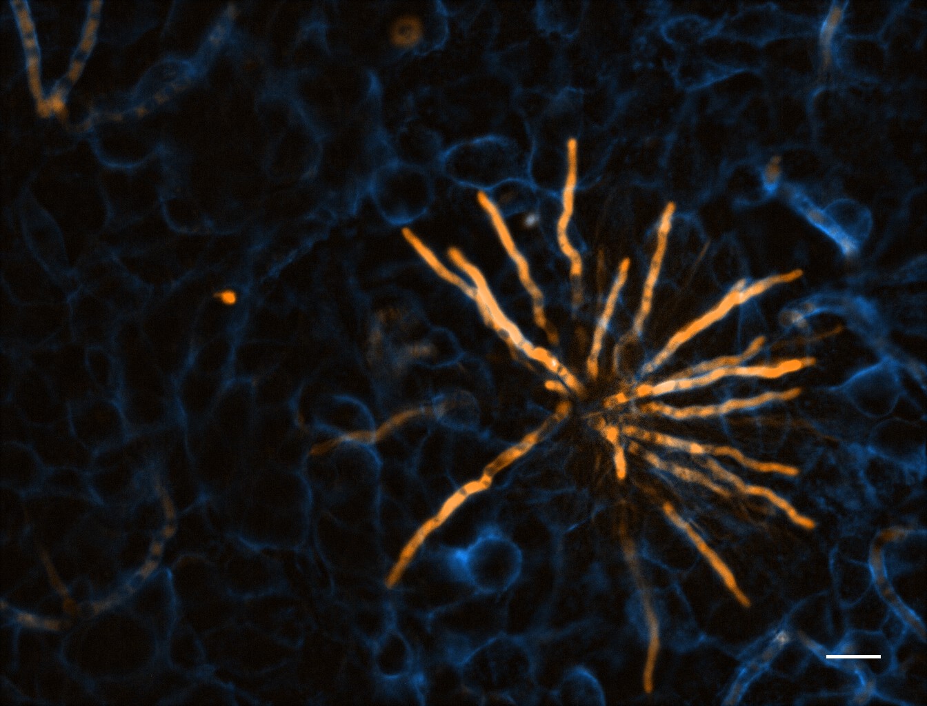
 Neurospora crassa hyphal filaments treated with fluorescently labelled siRNA (amber) to determine its localisation, with respect to nuclei (blue) and the fungal cell wall and septa (cyan). Scale bar = 5 µm. These experiments assessed the viability of siRNA as an anti-fungal treatment in pathogens. (Image courtesy of Ms Mireille Van Der Torre).
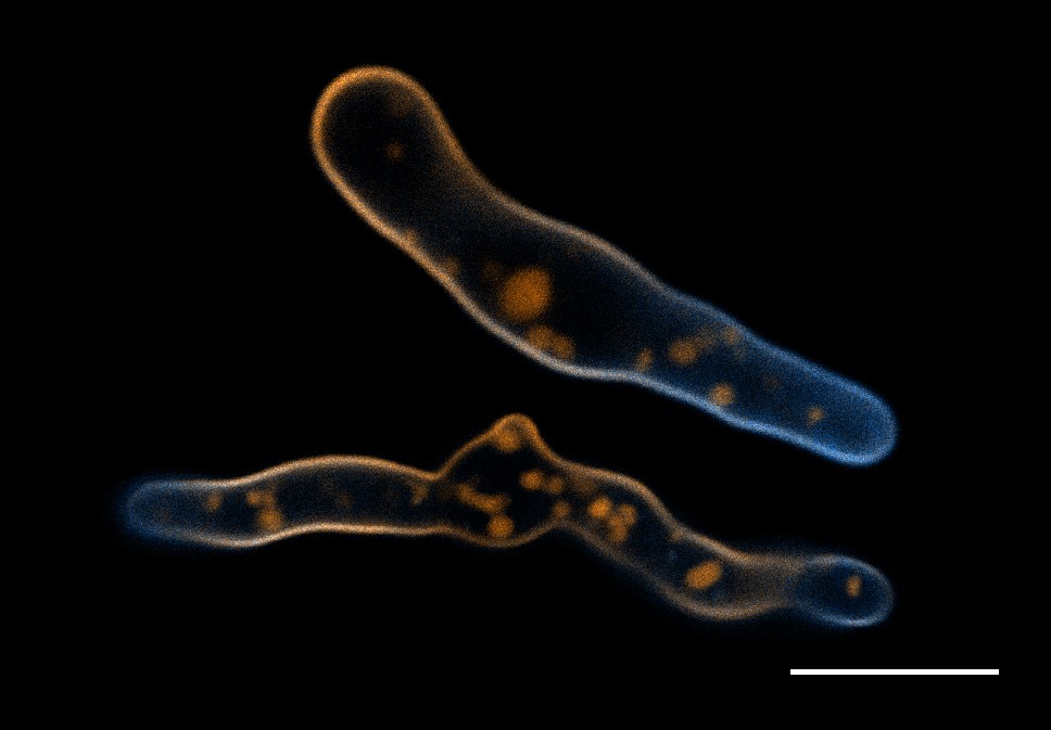 Aspergillus fumigatus germlings loaded with a synthetic cell membrane counterstain TMR-PAF96 (amber) and treated with fluorescent BODIPYcPAF26 anti-fungal peptide (cyan) to determine peptide’s localisation and mode of entry into the pathogen. Scale bar = 10 µm. (Image courtesy of Dr Can Zhao).
 TdTomato-expressing Aspergillus fumigatus (amber) microcolony after 12 hours development on an A549 epithelial alveolar monolayer labelled with ConA-FITC (blue). Scale bar = 10 µm. (Image courtesy of Dr Joy Icheoku).