LEDs Illuminate the Strathclyde Mesoscope
Gail McConnell (University of Strathclyde) and Brad Amos (University of Strathclyde and MRC Lab of Molecular Biology, Cambridge)
As soon as the confocal microscope came into wide use in the 1980’s, it revealed an inadequacy in the performance of microscope lenses. The optical sectioning effect which is the raison d’être of confocal optics simply did not occur with large specimens viewed in their entirety. The reason is that low-power microscope objectives have a very low numerical aperture, so the depth of field is large (of the order of 30 microns). For modern 3D microscopy this is useless, so we have designed a new type of objective lens which we call the Mesolens, because it has the wide field and large working distance of a macrophotography camera lens but the high resolution and good discrimination of depth of a medium-power microscope objective.
We have worked for several years with the lens designer and astronomer Esmond Reid to combine these properties in a novel optical design. The result, our x4 Mesolens, has an N.A. of 0.5, compared with 0.2 for a conventional objective of the same power. This produces a tenfold improvement in depth resolution and a factor of 2.5x in lateral resolution. Also, the lens is much faster (captures approximately six times more light) than its conventional counterpart. Unfortunately, such a lens is inevitably much larger than an ordinary objective and must be made to unusually tight manufacturing tolerances (Fig 1). Fig 2 shows how the Mesolens collects exquisite detail in a camera image from a field approximately 5mm in diameter. In Strathclyde, we have solved the problem of scanning with the very large beam sizes that the Mesolens needs and we have achieved confocal sectioning, scanning up to 20,000 lines per image instead of the usual 1000-2000.

Fig. 1. Professor Gail McConnell adjusting a specimen under the Mesolens objective in Strathclyde, watched by Dr Johanna Tragardh. The bulky central region of the lens houses the chromatic reflector.
The large (5mm diameter) field of the Mesolens means that LED sources are particularly well-suited to provide illumination. CoolLED Ltd and Chroma Inc. have collaborated with us to produce complex multi-band custom filters. Large-diameter chromatic reflectors and barrier filters have been developed which allow state-of-the-art high-intensity LEDs to be used, each with appropriate band-cleanup filters.
The University of Strathclyde, aided by a generous grant from the Medical Research Council to Professor Gail McConnell and a Leverhulme Emeritus Fellowship awarded to Dr Brad Amos, is now being equipped with Mesoscopes. In the ‘Mesolab’ (https://mesolab.wordpress.com) we have achieved our long-term goal: confocal imaging of mouse embryos at the 14-day stage, showing unprecedented subcellular resolution of all the organ systems. We are confident that this will be a game-changer in the study of mouse embryonic development, but the Mesolens is already proving its worth with diverse specimens. The Strathclyde lab has special expertise in nonlinear optics and since we know the entire lens prescription (normally kept secret by manufacturers), we have been able to calculate that two-photon excitation at certain wavelengths will be possible with the Mesolens and we will implement this soon, along with other novel imaging modes such as lightsheet.
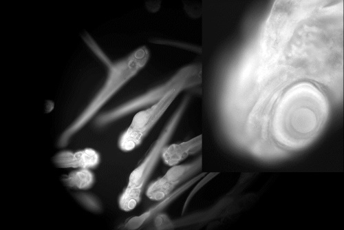
Fig. 2. Fluorescence image of 15 zebrafish larvae (fixed and stained with acridine orange) within the field of the Mesolens. A central portion of the image is shown enlarged at upper right: note the clarity of the retinal layers in the thin in-focus z level, which passes through the eye. This image shows how the Mesolens functions like a medium-power objective, but with 3mm working distance and a 5mm diameter field. The images could have sizes up to 400 Mb per channel per focal level.
