Growing a Bio-Imaging Network in North America
Meet the Experts

Alison North, Senior Director of the Bio-Imaging Resource Center, The Rockefeller University
Email: [email protected]
Tel: +1 212-327-7488
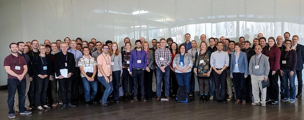
Alison North, Senior Director of the Bio-Imaging Resource Center at The Rockefeller University, and co-founder of Bioimaging North America (BINA), explains how the new network is bringing together core facility managers across North America.
Part of its aim is to help educate its members about new technologies, and we also spoke about Alison’s experience when it comes to LEDs as the go-to method for widefield illumination, and her advice on switching to this technology.
Can you tell us about you and your role?
I trained in Europe, last finishing in Manchester with a Wellcome Trust career development fellowship. When The Rockefeller University wrote to me and said they wanted someone to set up an imaging facility here, I only meant to stay for two years, and that was in 2000! I set up our imaging facility and since then it has grown enormously, and both people and the technologies are continually changing. All users have different challenges, different samples, different science and different needs; the challenge of teaching them is always different, and it never gets dull.
How did BINA get started?
The one thing those of us who founded BINA share is being part of national and international microscopy networks. We’ve all seen the value of this and realised it’s what the US community needs. What really started it for me was my favourite annual European Light Microscopy Initiative (ELMI) meetings and I attend these whenever possible even though I work in the USA. Over time I realised there was no equivalent meeting to network all US microscope core directors.
I make so many contacts at these meetings, whom I can ask if I have a question or need advice. The relationship between academia and industry is also unique at ELMI and it is viewed much more as a partnership. We attend company workshops to learn which technology is going to be useful, and this is important to us. The partnership between imaging scientists and companies is something that we really want to stress in BINA.

What is BINA and how can members get involved?
We want to help everyone get to know each other in different core facilities, bringing people together from remote corners of North America. Through BINA, we want people to exchange expertise and experience with different technologies and learn how to run a facility as effectively as possible.
We’re achieving this through working groups. One of the first groups we set up was the Quality Control & Data Management Working Group, as there is a huge push on including better metadata in papers and improving methods reporting so that experiments can be reproduced. We also want to promote good practices in microscopy and measure microscope performance, including measuring and comparing illumination intensities over time. Of course, that is one area where LEDs are coming out ahead.
A new Training & Education Working Group is where we will link to online material that will help people getting to grips with remote training. We’re encouraging everyone to send in links to YouTube videos or articles on everyday topics, for example we’ve just created one on how to focus a microscope without crashing into the slide. Another Working Group focuses on diversity and inclusion, and this will include helping individuals in remote geographical areas like Mexico. If we can get companies to offer advanced technical training, facility staff can undertake basic servicing themselves, instead of waiting for a service engineer to travel to Mexico.
We are also trying to secure funding for exchange visits between core facility directors and staff. After trialling these informally, it was amazing how many good ideas I got from visiting a few UK facilities. I’ve been in this job 20 years, but it’s always informative to hear how others run their facilities, and likewise, share my experiences.
More facilities are adopting LED illuminators; can you share your experience with this technology?
It started after I won an LED light source from a CoolLED competition and realised its simplicity and how easy it was to install. In theory we would like to avoid mercury lamps: I’ve had several explode over the years, so it’s always a concern. For a core facility manager, the convenience of LEDs is lovely, and it really cuts down on the amount of work needed to keep your instruments running.
LEDs have also really improved from the early days with their weakness in the green range, and now they provide more than enough light. Where I frequently work with live cells, we don’t run them anywhere near capacity. It’s such a bad idea to be exposing live cells to intense light and I think many scientists don’t quite understand that an image doesn’t need to be superbright to answer a biological question. In fact, I removed the fluorescence light source from my spinning disk system so that users couldn’t kill their samples by just looking for the cells! There might be times to turn the LED to maximum if you’re looking at fixed samples with weak staining, but I don’t think modern LED systems are at all limiting.
Despite the benefits of LEDs, one of the main challenges is justifying an alternative illumination source unless it has broken, or it is part of a new instrument. However, the upfront price point balances out if you consider the cost of mercury lamps and fibre optics over the next 10 years. Claire [Professor Claire Brown, Canada] found a ‘green’ lab initiative to fund switching over all of her systems, but I’m not aware of an equivalent scheme in the US. *
What advice would you share for updating a light source?
The main piece of advice I would give to somebody thinking of buying LEDs is to think through the whole system, particularly when it comes to selecting the right optical filters and especially if their dichroic mirrors are designed around lasers.
When we installed a 785 nm laser on the light sheet microscope several years ago, we needed a light source to visualise the dyes in that region, to check if stainings had worked. At the same time, users also need to check Alexa 350 dyes for the 405 nm laser. The LED light sources just becoming available at the time covered both ends of the spectrum, however optical filters were not widely available for these new LEDs. I received the best advice from Chroma, which was a really valuable piece of information I’d like to share: a lot of people don’t realise that many specialised filter sets have been designed that are not promoted on the website. Before purchasing a new light source, always speak to the light source manufacturer or directly to filter manufacturers such as Chroma, because you just don’t know what they have available.
Another point is that microscope companies generally supply standard illumination systems with the instrument, and this can sometimes be a mercurybased lamp like the metal halide systems. It’s a good idea when purchasing a new system to ask about other options such as LEDs.
*An increasing number of institutions are now offering ‘Green Grants’ to support the purchase of sustainable,
energy-efficient instruments such as LED Illumination Systems.
For more information on BINA, visit: www.bioimagingna.org
Learn how to select the best optical filters for LED Illumination Systems
