Validating LED illumination performance with Plug-and-Play confocal technology
Meet the Experts

Maria Giubettini, Application Specialist at CrestOptics
Tel: 0039.06.61660508
Email: [email protected]
Confocal microscopy can be an expensive upgrade for laboratories, but Dr Maria Giubettini, Application Specialist at CrestOptics explains a cost-effective technology for upgrading any widefield microscope.
Combining the CrestOptics X-Light V2 spinning disk and CoolLED pE-800 LED Illumination System enables both widefield and confocal on the same setup and is far more affordable than using laser illumination. Maria shares her recent experience validating the setup and what it means for life science researchers.
Can you tell us about your role at CrestOptics?
My background is in biology, where after my PhD I spent five years as a postdoctoral researcher. I was always fascinated by microscopy, so when I had the chance to move to CrestOpics, I took it immediately and I’ve been here as Application Specialist for three years.
We are based in Rome, Italy, and we develop and manufacture spinning disk confocal systems for fluorescence microscopy and diagnostic applications. I think the strength of our systems is flexibility and the ability to combine them with different devices to keep up to date with the latest technologies. It also means labs can start from the equipment they already own and gradually upgrade, always keeping up to date.
I’m involved in a variety of activities, from remote demonstrations to validating hardware configurations, including the setup with the X-Light V2 and CoolLED pE-800 Illumination System. I also support collaborations with researchers, where I might perform image acquisition and post-processing or provide advice and discuss the data, or how to perform quantification. It’s always exciting to provide technical support in this way and feel like I’m part of a research project. Staying in touch with the biomedical research field allows me to discover innovative applications and technologies and share the excitement of a new discovery. When I see an amazing image, or features that could previously not be resolved, it’s like a small discovery for myself too!
What are the main trends in this confocal technology?
When I look back to when I started with a wide field microscope 10 years ago and compare that to what’s available now, there are so many techniques evolving, with so many different microscopes and technologies; it really is impressive.
Many of our collaborating researchers would like to catch fast, dynamic events happening at the cellular level. This requirement for fast imaging fits perfectly with our spinning disk system since we can achieve fast image acquisition with high resolution and combine it with high throughput analysis. Another popular requirement we are becoming more involved in is super resolution imaging of thick specimens, such as organoids or even whole organs. I guess our technology is evolving to combine these different capabilities and obtain super resolution, even in thicker specimens.
This is where my background is useful to understand end user needs and explain to my colleagues what we need in order to satisfy a request. In fact, I think we can already satisfy many needs for these types of imaging to acquire high resolution data, required for more precise quantification and insightful information.
What are the benefits of using LED Illumination with your technology?
Every imaging facility ideally needs a variety of setups for different applications.
I think LED illumination technology is perfectly in line with our spinning disk features and advantages, such as low photobleaching or phototoxicity in live experiments.
The LED technology also presents an optimal solution for fixed samples, where our system allows high efficiency of light passing through the optics. The combination can be suitable for many different applications. Last but not least, LEDs offer a cost-effective advantage in terms of the balance between the power and price.
I have worked with many different light sources in combination with different microscope setups, and personally I have had a positive experience with LED illumination.
How did the CrestOptics X-Light V2 and CoolLED pE-800 perform?
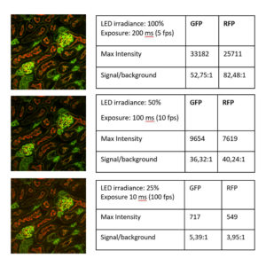
I had the chance to test this configuration using different samples including tissue sections, and it really exceeded my expectations. I obtained high-contrast images (Figure 1), even with low irradiance and short exposure times. I’ve been impressed by the improvement of LED illumination with the CoolLED pE-800, and especially in combination with our confocal technology.
The data demonstrated without a doubt how the CoolLED pE-800 and X-Light V2 represent an excellent combination at a reasonable price. This setup allows researchers to obtain high optical sectioning power and this is the case with many different samples, from brighter to dimmer, and the ability to optimise the setup for the sample is a plus point.
How have you found working with CoolLED?
I have really enjoyed our collaboration so far and appreciate our discussions and sharing ideas – and of course achieving excellent scientific results. I hope we can meet one day and get together in front of a microscope!

Figure 1: Validating the CrestOptics X-Light V2 and eight-channel CoolLED pE-800 LED Illumination System. A range of LED irradiance settings and exposure times demonstrated high-contrast data, even at 25% irradiance and 10 ms exposure. Fixed mouse kidney stained with GFP and RFP. Plan Apo Lambda 60X Oil objective; captured using Photometrics Prime 95B camera. Images and data courtesy of CrestOptics.
