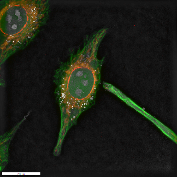
Fixed HeLa cells stained with Antibody for cytoskeleton (green), mitochondria (orange) and nuclear membrane(red). The whole cell was also imaged with Nanolive’s holotomographic technology and the four images were overlapped to create the overlay. Image courtesy of Nanolive.
More information available here: www.nanolive.ch/products/3d-microscopes/fluo

