Authors
Ya-Ling Tan et al.
Topic
Molecular Biology
Extract
"Acquire fluorescence images of the entire microwells using an inverted confocal laser scanning microscope under 10× objective.
Preparing. Place the droplet counting microwell chip on the microscope counter. Open the imaging software and select appropriate scan modes.
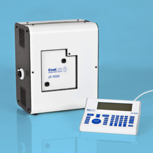
Note: Nikon Inverted Eclipse Ti epifluorescence microscope equipped with a motorized XY stage (Nikon), a CoolLed pE-4000 illumination source, a Nikon DS-Qi2 camera, and an apochromatic 10× objective (numerical aperture, 0.45) (Nikon) is used here."
Product Type
Journal
Star Protocols
Year of Publication
2022

