Where fluorescence and label-free imaging meet
Meet the Experts

Mathieu Frechin, Head of Quantitative Biology, Nanolive
Email: [email protected]
Tel: +41 21 353 0600
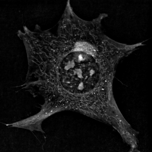
During live cell imaging experiments, it can be a challenge to ensure fluorescence is not interfering with the biological processes you wish to observe. For studying cellular structures, label-free imaging technology created by Nanolive overcomes this. To combine high quality holotomographic data with molecular data from fluorescent markers, one Nanolive system is integrated with a CoolLED Illumination System based on the pE-300ultra. Head of Quantitative Biology at Nanolive, Mathieu, explains the technology in more detail and gives an insight into life at Nanolive.
Can you tell us about Nanolive and your role?
Here at Nanolive, my role as Head of Quantitative Biology covers a large spectrum of responsibilities. When I joined the company in 2017, I built the cell biology lab and developed post-acquisition and computer vision techniques for cell biology. It is very multi-disciplinary which suits my background in molecular cell biology and biophysics. In particular, I studied the biology of mammalian cell membranes which is helpful given that our microscope is able to image membranes in great detail. Label-free imaging is a dynamic field, and our technology was created after a breakthrough discovery made by Dr Yann Cotte (Nanolive’s CEO) and Dr Sebastien Equis (CTO). The concept of holotomography itself is not new, but how the approach was improved and implemented is. As far as I’m aware, we are the only platform in the world to reach such high levels of performance using this approach.
What are the main applications of your technology?
The main application is live cell biology. Most scientists are interested in the microscope because of its label-free aspect, as its capacity to observe organelles is very good. This includes mitochondria and lipid droplets; major components of cellular metabolism. Its low phototoxicity also makes it popular for scientists working with stem cells. Low phototoxicity means you can image for extended periods of time and even weeks, without harming the sample.
Why is label-free imaging becoming so popular?
There are three clear advantages: Firstly, you do not have labels that interfere with the phenomenon you want to observe. Secondly, you don’t have phototoxicity because we work at levels of light that are three to four orders of magnitude lower than standard widefield fluorescence excitation. One of the big reasons why we are successful is that more and more scientists want to make sure their images are artefact-free. The third advantage lies in the multiplexing aspect: when you work label-free, you acquire data on many different biological processes at once, making images extremely rich. This means they are also complex and require advanced computer vision solutions. Another advantage is the convenience of a short sample preparation time and easy acquisition.

How are label-free and fluorescence microscopy complementary?
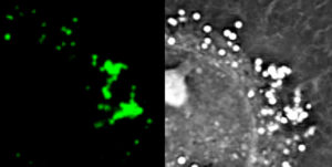
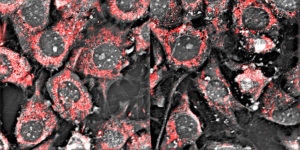
Holotomography looks at membranes, not molecules, so there will always be a requirement for fluorescence microscopy when scientists want to localise molecules. We need to take the best of both worlds. For example, identifying organelles through fluorescence and monitoring their structures and dynamics non-invasively.
What do you need from a fluorescence light source?
Number one is reliability, as we don’t want the systems being returned. Like every microscope, we also need uniform illumination. With our CoolLED Illumination System, we are happy with both these factors. There are many more positives: quality and price, simplicity and robustness and flexibility. But it’s more than that, on the company side everyone we speak to from CoolLED is always kind and helpful.


Why did you choose to work with CoolLED?
After meeting at ASCB in winter 2016, we received a CoolLED Illumination System prototype which was integrated and tested over February 2017. The first microscope with a CoolLED Illumination System was sold just later that year. In addition to meeting technical specifications, CoolLED was very quick to react to changes in our situation and provide appropriate solutions.
This all took place before I joined Nanolive, but I was already familiar with CoolLED from my postdoc where I was responsible for the imaging equipment in my team. CoolLED has always been my favourite light source. It’s less expensive and easier to control than lamps which can be very tedious to work with.
Where is the future of labelfree imaging?
Our microscopes create such a wealth of data, the future is really in developing applications that allow our customers to harvest that data in a meaningful and simple way. The major challenge is to integrate Artificial Intelligence (AI) into image analysis. To me, that is where the future lies.
Discover label-free imaging at: nanolive.ch
