Becoming Mercury-Free with the ‘Image in an Image’ Competition
Meet the Experts

Alfonso Schmidt, Bioimaging Specialist, Hugh Green Cytometry Centre, Malaghan Institute of Medical Research
Tel: +64 4 499 6914
Email: [email protected]
We recently started running an imaging competition with a difference, where entrants submit a captivating light microscopy image which contains another image (confused? Find out more here).
Winner of the 2020 Image in an Image competition was Alfonso Schmidt, Senior Staff Scientist and Bioimaging Specialist at the Malaghan Institute of Medical Research, Hugh Green Cytometry Centre in New Zealand. He explains how he discovered his ‘Eye of Sauron’ image and how the prize – a pE-300ultra Illumination System – is helping the facility to become mercury-free and make breakthrough discoveries in immunology.
Tell me about yourself and your role
I’m originally from Chile, South America and moved to New Zealand in 2010. I have been working at the Malaghan Institute of Medical Research for the past 10 years, where I am a senior staff scientist and bioimaging specialist. The Institute focuses on specific questions related to immunology in the context of different diseases such as asthma, allergy, cancer, gut health, brain health and infectious disease.
I manage the histology and bioimaging core, part of the Hugh Green Cytometry Centre, which provides support and training in flow cytometry, cell sorting and genomics, as well as microscopy for the scientists at the Malaghan Institute and other scientific organisations.
I’m responsible for providing the best microscopy service and advice as it relates to a scientist’s experiment. Most researchers know what they want but might not have the expertise or knowledge to make optimal use of the tools available to them to make that happen. As a support scientist, it is important for us to provide not only the options, but recommendations to improve upon existing experiments. For example, one kind of microscopy technology may provide higher image resolution, but another might offer higher sample throughput. I enjoy having these kinds of discussions with our researchers, opening their eyes in terms of the technology we have and what the best solution is for them.
I’m really passionate about microscopy and have always liked photography, so in a way I’m like a micro-photographer, and this is how I explain my job to my kids. I think one of the most enjoyable aspects of microscopy is the ‘wow’ factor it brings. Compared to something like a graph plot showing data points, there is something about a microscopy image that amazes people and captures their imagination.
How did you choose your competition entry?
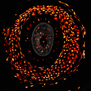
If time allows, I always try and save at least one or two additional images from each experiment, whether they end up in a scientific report or not. Images that provide the best experimental data are not necessarily the most beautiful, and the most ‘artistic’ images might not convey as much scientific meaning, but that doesn’t mean they don’t have value. The ‘Eye of Sauron’ was one of these artistic images, from a study of atopic dermatitis (eczema) which is an allergic reaction causing a rash and dry skin. I noticed the interesting arrangement of the cells around the hair follicles at the time. Not long later I heard about this competition and this image naturally seemed to fit.
All we needed was to come up with a name. At the time, I had been watching the Lord of the Rings movies with my family, and I recalled how much it looked like the Eye of Sauron. As the movies were filmed here in New Zealand, it felt like the name represented where we are too.

How has the new light source helped your facility?
It has been a great privilege to win the ‘Image in an Image’ competition, and we are really enjoying the pE-300ultra. Updating our epi-illumination light sources is one of the best ways to upgrade our imaging facility and provide better tools for better science.
The pE-300ultra has also brought us closer to removing mercury completely from our facility. Organisations throughout New Zealand are concerned about safe working environments, and mercury is of course a toxic chemical. Previously we had old-fashioned mercury lamps, which only lasted 300 hours and were frustrating to replace and dispose of because of the mercury. We had just upgraded our stereo and inverted microscopes with CoolLED pE-300white Illumination Systems, and now with the additional pE-300ultra we are almost completely mercury-free.
The CoolLED Illumination Systems are fantastic, and I really want to highlight how easy they are to use, which is great from a training perspective. In terms of microscopy, the flexibility of selecting and tuning individual channels is beneficial to prevent photobleaching. LED microscope lighting has improved so much in the last few years, and every layer of extra capability makes the microscope setup more interesting to use.
Winning the competition has also been really rewarding for me personally and for the wider Institute – we’re a small facility in a little-known area, but we do some really fantastic work!
What research has the pE-300ultra been used for so far?
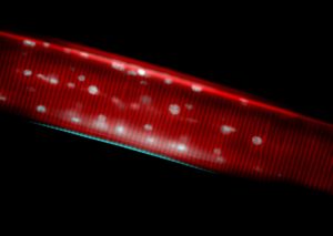
The pE-300ultra Illumination System has been fitted to our Olympus BX51 upright microscope, which currently is mainly used by a research group working with hookworms as a therapy for autoimmune conditions such as inflammatory bowel disease. We are using this microscope setup to monitor how the worms develop by quantifying cell nuclei over time using several culture conditions.
This work, which is currently in clinical trials, is based on the evidence that certain species of human hookworm may play a role in ‘dampening down’ harmful inflammation in the body. Hookworms have evolved over millions of years to manipulate their hosts in order to survive, altering the human immune system to evade detection and expulsion. In places that have adopted modern hygiene practices, we see a corresponding rise in allergic and autoimmune disorders. The Malaghan Institute has initiated this clinical programme, in part to explore whether there is therapeutic potential of human hookworms for managing these conditions. Our aim is to ultimately find better treatment options for a range of inflammatory and autoimmune diseases, including coeliac, asthma, allergy, MS and inflammatory bowel disease.

