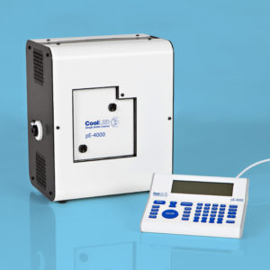
Time series of a confluent sheet of keratinocytes migrating during a live-cell imaging outgrowth assay, acquired on an Olympus IX83 microscope equipped with a CoolLED pE-4000 Illumination System. Scale bar = 150 μm. Image courtesy of Graham Wright, Sarah Zulkifli, Tan Tong San, Declan Lunny, John Common & Birgitte Lane
Read the application note: Applying microscopy to understand skin fragility disorders

