


Overview
Resulting from our collaboration with the University of Strathclyde, the pE-340fura is a bespoke LED Illuminator for Fura-2 ratiometric calcium imaging. The calcium imaging Light Source utilises the successful pE-300 Series platform, and also supports everyday fluorescence microscopy in a compact and affordable package.

The 340 nm and 380 nm LED illumination system provides the optimum excitation wavelengths for Fura-2-based calcium imaging, allowing high-precision, stable, high-throughput imaging with video-rate time resolution.
High-speed acquisition
Until recently, the response time of illumination systems for Fura-2 imaging has been limited to milliseconds due to mechanical switching of the wavelengths in arc lamp and monochromator systems. However, the new pE-340fura can be controlled via convenient BNC TTL connections for precise illumination control in as little as 20 microseconds.
Improved cell viability and cost
Using the new pE-340fura LED Illumination System, less Fura-2 dye can be loaded into the cells whilst still maintaining the same measured calcium concentration and good signal-to-noise ratio. The reduction in required dye not only improves cell-viability due to reduced dye toxicity, but also results in a cost reduction per experiment.
Improved signal to noise
Work by Sandrine Prost et al., from the University of Edinburgh, has shown that with independent wavelength controllable LED sources, signal-to-noise is dramatically improved over bulb systems and even over some available white broad-spectrum LED sources.
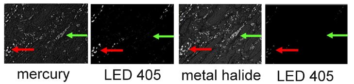
High levels of autofluorescence and fast photobleaching of specific fluorescence when illuminating Qdots with Metal Halide.2
- Direct or light guide delivery options – flexibility of UV optimised attachment options
340 nm Excitation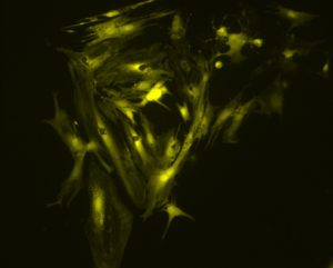 |
380 nm Excitation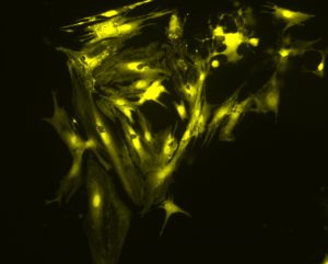 |
Figure 1: cardiac myocytes. The cells were loaded with Fura-2 using standard conditions (incubation with 2 mM Fura-2 acetoxymethyl ester for 30 minutes, followed by an additional 30 minutes for de-esterification). Images were obtained by Martin Bootman and Katja Rietdorf, School of Life, Health and Chemical Sciences, The Open University, UK.
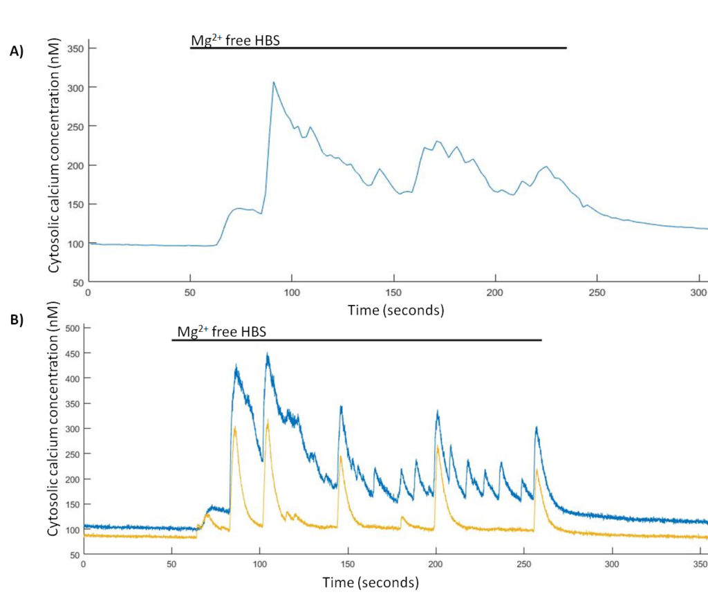
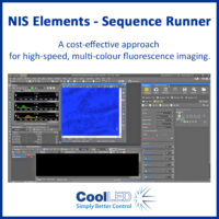
Spontaneous Ca2+ events are induced in Mg2+-free HBS. (A) Representative trace from a single hippocampal neuron of Mg2+-free induced Ca2+ events imaged at 0.5 Hz and (B) representative trace from two hippocampal neurons of Mg2+-free induced Ca2+ events imaged at 24.39 Hz.1

Comparison of Ca2+ increases obtained from the application of trypsin (100 nM) to tsA-201 cells loaded with different concentrations of Fura-2 AM.1
References
- TINNING, P. W., FRANSSEN, A. J. P.M., HRIDI, S. U., BUSHELL, T. J. and MCCONNELL, G. (2017), A 340/380 nm light-emitting diode illuminator for Fura-2 AM ratiometric Ca2+imaging of live cells with better than 5 nM precision. Journal of Microscopy. doi:10.1111/jmi.12616
- Prost S et al (2016) Choice of Illumination System & Fluorophore for Multiplex Immunofluorescence on FFPE Tissue Sections. PLoS ONE 11(9): e0162419. doi:10.1371/journal.pone.0162419
Performance
- Dedicated 340 nm and 380 nm outputs for Fura-2 ratiometric calcium imaging
- Broad white output (435 nm – 645 nm) for imaging standard fluorophores
- Microsecond switching (340/380) for capturing high-speed events
- Stable LED illumination minimises noise below that of experiment, reducing false positives and increasing precision.
- Higher signal-to-noise gives cleaner images and data whilst requiring less Fura-2 dye, reducing toxicity and costs
- UV optimised optics delivers maximum irradiance on standard microscope configurations
- Individual channel triggering via TTL in microseconds
- Removable inline excitation filter holders: no moving parts for fast acquisition
- Sequence Runner – sequenced excitation from a single TTL-out
- Individual channel selection controls the level of excitation of each dye independently on a multi-stained sample
- Compatible with most imaging software for integrated control with imaging set up
- Precise irradiance control in 1% steps (0-100%): no ND filters required
Convenience
- Specify for existing single and multi-band filter sets: no need to buy new filters
- Simple to fit, simple to use: no alignment, a once only adjustment
- Wide range of microscope adaptors: fits most microscopes
Plus all the benefits of LED technology
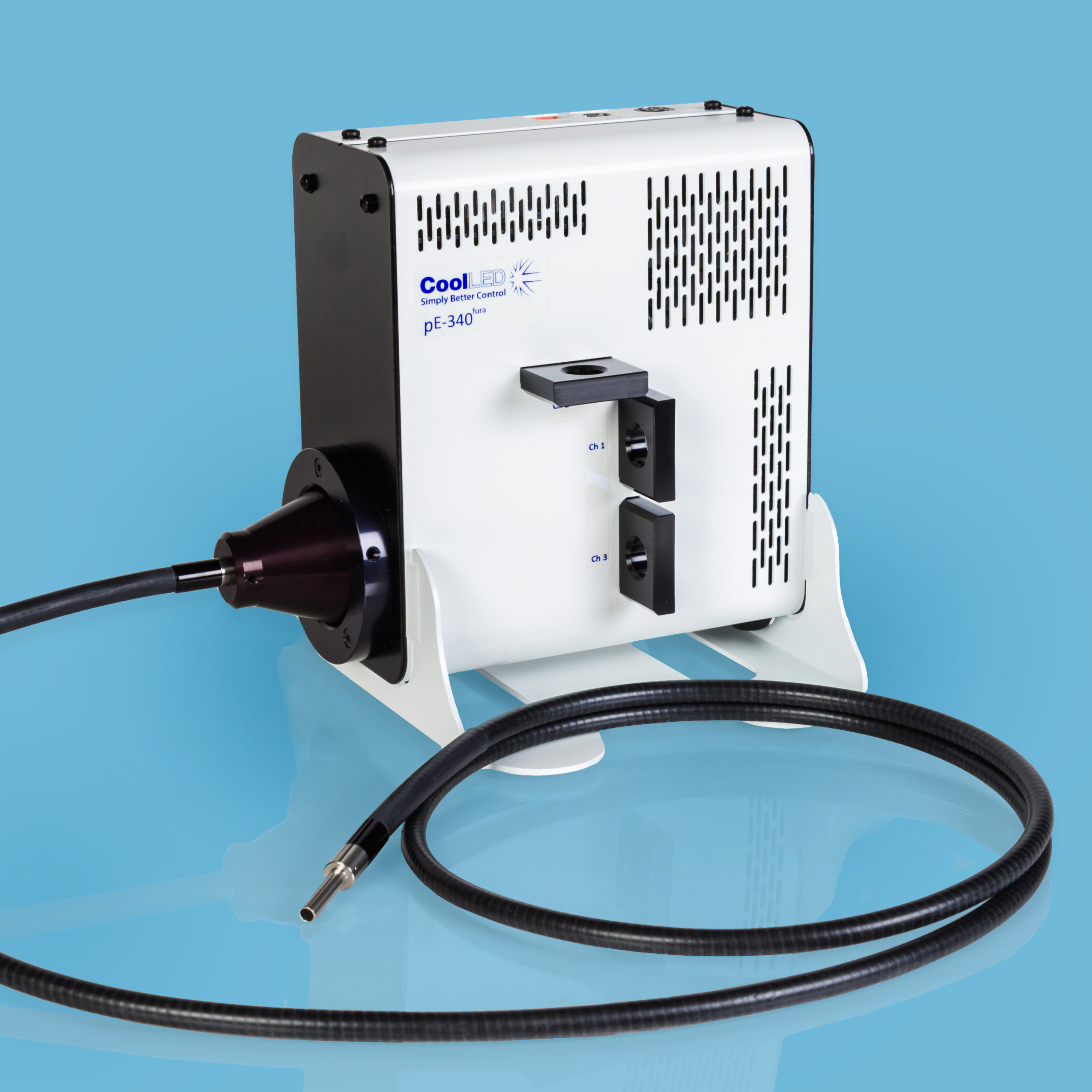
The system comprises a pE-340fura Light Source, Control Pod, set of three Excitation Filter Holders and Power Supply.
Control
Control pod for operation and access to settings and usage history.
Manual:
Manual control for instant on/off and irradiance control in 1% steps from 0 – 100%
Individual bands can be selected/deselected and controlled independently as desired
Remote:
Via global and individual channel TTL for on/off control of selected channels using a BNC connection on the light source.
Triggering speed <20 µs at full power
Connectivity:
Remote via USB (B type) for imaging software control (see Imaging Software)
Sequence Runner:
Single TTL input to step through user defined sequence.
Triggering speed <20 µs at full power
Choosing configurations
There are two pE-340fura‑configuration options:
Direct-fit: Higher irradiance is possible when directly connecting to a microscope. Select from a range of microscope adaptors which covers all current and most older models. A simple once-only adjustment will allow optimisation to the optical path of the microscope.
Liquid Light Guide: If there is a need to keep the source of illumination remote from the microscope, the Liquid Light Guide variant is available for use with a 3 mm diameter Liquid Light Guide. An optional pE-340fura Universal Collimator can be specified in conjunction with a microscope adaptor if required, containing optics optimised to transmit the 340 nm. We now include a light source stand for the pE-340fura Illumination System ordered for light guide delivery.
Specifications
Power requirements
100-240 V a.c. 50/60 Hz
Power consumption
Standby Max 2 W
1 band (white) at 100% irradiance Max 24 W
2 bands (340 + white) at 100% irradiance Max 30 W
3 bands (All) at 100% irradiance 36 W
Dimensions
pE-340fura Light Source: 77 mm(w) x 186 mm(d) x 162 mm(h) – Weight 1.40 kg
pE-340fura Control Pod: 88 mm(w) x 125 mm(d) x 37 mm(h) – Weight 0.32 kg
pE-340fura Power Supply: 167 mm(w) x 67 m(d) x 35 mm(h) – Weight 0.62 kg
pE-Universal Collimator: 44 mm(w) x 86 mm(d) x 44 mm(h) – Weight 0.17 kg
Environment and Safety
- Mercury-free and Laser-free
- Energy Efficient
- Long lifetime
- No bulb replacements
- Reduced risk of eye damage
- Quiet operation
- No special disposal regulations or issues
Warranty
Industry-leading 36 months (please note, the 340 nm LED is warranted for 3000 hours accumulated use). Read more here.
All data correct at time of publication

We are often asked about the power, intensity or irradiance of CoolLED Illumination Systems. The answer is not as simple as you might believe from many websites and data sheets, and it can be difficult to compare data from these sources as measuring set ups differ.
Photons are also lost as light travels from the light source to sample plane. The only way to objectively compare light sources is by measuring irradiance at the sample plane. To understand why we use the term irradiance and how to measure and compare light sources with accuracy and precision, download our white paper. Or please contact us if you have further questions.
For performance data, please contact us
The system is very useful and easy to use, which makes it much more accessible for everyone who is interested in ratiometric calcium measurements.
Dr Peter Szentesi, Research Associate, Department of Physiology, University of Debrecen (pE-340fura)
The CoolLED pE-340fura system with the 340 and 380 nm wavelengths and white light: Number one I was really surprised how bright the white light was, which is really useful for all other types of imaging. Number two, the 340 and 380 nm wavelengths are really well implemented with the excitation filters that match the filter cube in the microscope that you can just slide into the CoolLED system and then adjust the intensity of the 380 nm to balance that out for Fura-2 experiments. It’s really super easy.
Ken Anderson, BioVision Technologies (pE-340fura)
I can absolutely recommend this light source for any kind of Fura-2 measurements.
Lars Kaestner and Quinghai Tian, Molecular Cell Biology, University of Saarland, Germany (pE-340fura)
Research Papers
- Up-regulated expression of two-pore domain K+ channels, KCNK1 and KCNK2, is involved in the proliferation and migration of pulmonary arterial smooth muscle cells in pulmonary arterial hypertension
- Genetically engineered HEK cells as a valuable tool for studying electroporation in excitable cells
- PINK1 and Parkin regulate IP3R-mediated ER calcium release
- Rescue of astrocyte activity by the calcium sensor STIM1 restores long-term synaptic plasticity in female mice modelling Alzheimer’s disease
- Oxoglutarate dehydrogenase complex controls glutamate-mediated neuronal death
White Papers
- Cell Biology in the Fast Lane: Analysing Fast Cellular Processes with LED Microscopy Illumination (pE-800fura)
- Revealing brain function with intracellular calcium imaging and patch-clamp recordings
- Understanding mitochondrial stress self defence
- Particulate matter-associated Ca2+ signalling and modulators
Sequence Runner for Nikon NIS Elements
Sequence Runner: Cost-effective automation
CoolLED Multi-Wavelength LED Microscopy Illumination
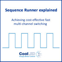
Sequence Runner for high-speed imaging
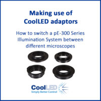
Making use of CoolLED adaptors
Configuring cellSens

Configuring LAS X
Configuring Hamamatsu HCImage
Configuring MetaMorph

A 340/380nm LED Illuminator for High Precision Fura-2 Ca2+ Imaging

An Introduction to Calcium Imaging
| Light Source | Application | Type | CoolLED LED | Chroma | Semrock | |||
|---|---|---|---|---|---|---|---|---|
| pE-340fura (Direct/LLG) | Fluorescence | Single | 380 | 39000 AT Dapi | 49028 ET Dapi 395 | LED-DAPI-B | ||
| pE-340fura (Direct/LLG) | Fluorescence | Single | Blu/Grn (410-750) | 49001 ET eCFP | 49013 ET Teal | LED-CFP-A | LED-mTFP-A | |
| pE-340fura (Direct/LLG) | Fluorescence | Single | Blu/Grn (410-750) | 49054 CoolLED GFP | 49002 ET eGFP | GFP-4050B | GFP-A-Basic | |
| pE-340fura (Direct/LLG) | Fluorescence | Single | Blu/Grn (410-750) | 49303 ET Green#1 | 49003 ET eYFP | |||
| pE-340fura (Direct/LLG) | Fluorescence | Single | Blu/Grn (410-750) | 49004 ET Cy3 | 49005 ET dsRed | TxRed-4040C | LED-TRITC-A | |
| pE-340fura (Direct/LLG) | Fluorescence | Single | Blu/Grn (410-750) | 49055 CoolLED mCherry #1 | 49055 CoolLED mCherry #2 | |||
| pE-340fura (Direct/LLG) | Fluorescence | Single | Blu/Grn (410-750) | 49008 ET mCherry | 39010 AT Tex. Red | YFP-2427B | LED-mCherry-A | |
| pE-340fura (Direct/LLG) | Fluorescence | Single | Blu/Grn (410-750) | 49006 ET CY5 | 39007 AT Cy5 | Cy5-4040C | ||
| pE-340fura (Direct/LLG) | Fluorescence | Pinkel | 340, 380 | T400lp & ET510/80m | FF409-Di03 | FF01-510/84 | ||
| pE-340fura (Direct/LLG) | Fluorescence | Pinkel | 340, 380, White (BLU/GRN) | T495lpxru, ET470/40x, ET535/70m | ||||
| pE-340fura (Direct/LLG) | Fluorescence | Pinkel | 340, 380, White (BLU/GRN) | 79003bs, 79003m, ET555/25x | ||||
| pE-340-FR-D-YYY-ZZ: | pE-340fura Illumination System. Direct Fit. Includes Light Source, exchangeable microscope adaptor (YYY) to customer-specified microscope, remote manual control pod, set of three excitation filter holders (25mm diameter), Excitation Filters for 340 nm & 380 nm, and power supply (ZZ). |
| pE-340-FR-L-ZZ: | pE-340fura Illumination System. For use with 3 mm Liquid Light Guide. Includes Light Source, stand, remote manual control pod, set of three excitation filter holders (25 mm diameter), Excitation Filters for 340 nm and 380 nm, and power supply (ZZ). |
| pE-1906:
pE-1908: |
1.5 m long, 3 mm diameter liquid light guide
3 m long, 3 mm diameter liquid light guide |
| pE-340-FR-COLL-YYY | pE-340fura Universal Collimator for use with a single liquid light guide. Includes Microscope Adaptor (YYY) to customer-specified microscope |
To specify microscope code (YYY) see Adaptors
To specify power cable (ZZ): 10 = Australia, 20 = Europe, 30 = UK, 40 = USA
More information is available on the Accessories tab.
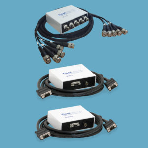
USB-TTL Conversion kit
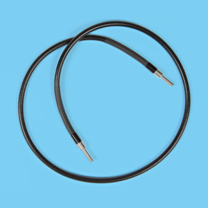
Liquid Light Guide
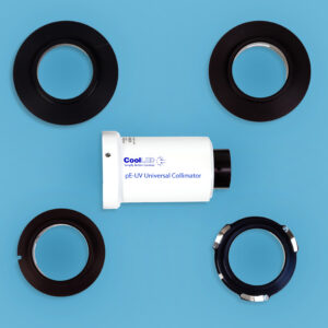
pE-UV Universal Collimator
The CoolLED pE Driver is first required for software integration, and can be downloaded here.
Contact CoolLED for further information.
